Corneal cross linking
Corneal cross-linking is a minimally invasive outpatient procedure designed to treat progressive keratoconus (and, sometimes, other conditions that cause a similar weakening of the cornea).
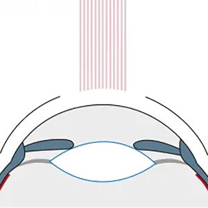
The corneal cross-linking procedure strengthens and stabilizes the cornea by creating new links between collagen fibres within the cornea. The two-step procedure applies specialty formulated riboflavin (vitamin B) eye drops to the surface of the eye immediately followed by a controlled exposure of the eye to ultraviolet light.

Best candidates for corneal cross-linking
Corneal cross-linking is most effective if it can be performed before the cornea has become too irregular in shape or there is significant vision loss from keratoconus. If applied early, cross-linking typically will stabilize or even improve the shape of the cornea, resulting in better visual acuity and an improved ability to wear contact lenses.
What to expect during and after corneal cross-linking
During the initial consultation your eye surgeon will measure the thickness of your cornea and make sure you are a good candidate for the procedure. You also will need to have a compressive eye exam to assess your vision and general eye health.
Detailed mapping of the shape of your cornea (called corneal topography) will also be performed.
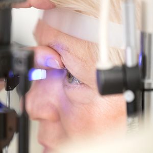
The cross-linking procedure takes about an hour in most cases. During this procedure the eye are fully numbed with anaesthetic eye drops. Following this, the first layer of the cornea (epithelium) will be removed and the vitamin B eye drops are installed every 2 minutes for 30 minutes. After this a special UV light is applied to the cornea for 8 minutes which is a new advanced technique called rapid cross linking.
Your Personalised Procedure
To determine which procedure is right for you, a comprehensive assessment is conducted utilizing state-of-the-art technology. Based on the thorough diagnostic assessment, your surgeon will tailor a treatment plan to match the right procedure for your unique vision needs.


